
Artifacts
Dr. Luca Bartalini, TSRM is a renowned Italian MR Imaging Specialist and Consultant with over 10 years of additional experience in multi-modal clinical imaging.
Currently a consultant to Esaote – Dr. Bartalini also happens to be an expert in MR artifact identification and associated corrective techniques. He is the author of the highly acclaimed imaging text – “Artifacts and Techical Solutions in MR Diagnostic Imaging”
Around October – November 2019, We, the makers of MRI Buzz – Matt Rederer BS RT (R) (MR) (CT) MRSO MRSE (MRSC) and Lubna Baig (Lexie) BSRT (R) (MR) – reached out to Dr. Bartalini with a request to collaborate with us and oversee the artifacts section of our site – a position he was gracious enough to accept.
With his help; students, aficionados, and patients of MR/MR Safety/Diagnostic Medicine, alike, will be able to add to their understanding of MR imaging artifacts and relevant/proposed technical solutions.
Common Artifacts
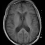
This artifact is a result of undersampling in the phase direction.
Direction: Phase
Remedy
- Swap Phase Direction
- Increase field of view/FOV
- Utilize no phase wrap/phase oversampling
- Use saturation bands over wrapping tissue
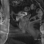
This artifact is seen where tissues with different magnetic susceptibility are experienced (Ex. Air=Paramagnetic and Tissue=Diamagnetic)
Direction: Slice
Remedy
- Scan on a lower field strength when possible
- Increase Receiving bandwidth/rBW
- Increase Frequency encoding
- Thinner Slices
Patient imaging occurs at isocenter. This is the area where we have a uniform magnetic field and can predict hydrogen behavior. When we image outside that area, tissues may not be represented appropriately.
Direction: Slice
Remedy
- Decreasing the FOV
- Avoiding techniques sensitive to homogeneous fields such as gradient echoes and fat saturation techniques.
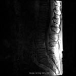
When slices overlap, overlapping tissue will experience multiple excitation pulses, this will result in suppression of those tissues.
Direction: Slice
Remedy
- Angling overlapping slices so overlapping is minimized.
- Use multiple scans to image area (Ex. Axial T2 through L4-5, then Axial T2 through L3-4. etc.)
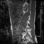
When slices are too thick, they can start to average many different tissue types together blurring our image.
Direction: Slice
Remedy
- To reduce this artifact, we can use thinner slices.
This artifact is a result of a tissues permittivity. Depending on the type of tissue and the thickness of that tissue, we can experience a loss of signal in part of our image.
Direction: Slice
Remedy
- Utilize dielectric pads
- Use filling
When we have a corrupted pixel in our k space, we can produce a pattern effect in our image. We can actually predict where the corrupted pixel is by reviewing the pattern produced.
Direction: Slice
Remedy
- This artifact usually goes away when repeating the sequence. If it does not, a service engineer should be contacted.

This is an artifact produced when RF contamination occurs when data is collected.
Direction: Frequency
Remedy:
- Check seal on scanner door for leaks
- Check for equipment contamination in the scan room
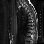
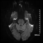
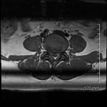
This artifact is commonly seen with spine and knee imaging
when multichannel surface coils, phased array coils, and fast pulse sequences are used. We typically see this artifact on sagittal and coronal planes. When interference is detected from structures outside the FOV, this artifact is seen and is considered a technical error.
Direction: Phase
Remedy: Check coil selection
anatomical structure being scanned antenna.