MR Safety Education

Quick Jump To:

Level 1 MR Personnel Training

This video provides basic information for individuals who require training that encompasses MR safety knowledge for individuals that may find themselves in the MRI environment and can care for themselves, not patients or personnel. It is not meant to be a complete educational video, just a quick annual refresher.

[ulp-enroll-course id=”36337″]

Please enroll and take the post quiz in this course to earn your certificate.

Find the course Here

Level 2 MR Personnel Training

This video provides basic information for individuals who require training that encompasses MR safety knowledge for individuals responsible for the care of their patients and other personnel in the MR environment. It is not meant to be a complete educational video, just a quick annual refresher activity.

[ulp-enroll-course id=”36332″]

Please enroll and take the post quiz in this course to earn your certificate.

Find the course Here

Reading manufacturer Information

Static Magnetic Field

When identifying the limits associated with the static magnetic field, we need to understand the following statements:

This means that we can scan at only 1.5T and no other field strength.

This means that we can scan at any field strength except 3.0T or greater.

This means that we scan scan our patient at a 1.5T.

1.5T = 64MHz

3.0T = 128MHz

This refers to the magnetic spatial gradient. A magnetic spatial gradient plot on the MRI unit you are using should be reviewed to see if this limit can be met.

Time-varying RF

When identifying the limits associated with the time-varying RF field, we need to understand the following statements:

The modes of operation are dependent on the type of RF coil we are using. We could be using a whole body RF coil (the one inside the gantry) or a local RF coil (one placed surrounding the body part).

Normal mode whole body is limited to 2W/kg except for the head which is 3.2W/kg.

This refers to the estimated dose of RF energy absorbed by our patient. It is measured in W/kg and can be associated with the IEC 60601-2-33 operation modes.

This refers to the accurate dose of RF energy to a tissue. The B1 refers to hte RF field and the “+” refers to the energy yeilding component of the RF. The root mean square refers to an average over a period of time. Every system should have an area that displays the B1+rms

Time-varying gradients

When identifying the time-varying Gradient fields, we need to understand the following statements:

The modes of operation control the time-varying magnetic fields. This refers to both the RF and Gradient fields. We discussed the RF fields, but normal operation mode is meant to not cause physiological stress to our patient. 

In regards to the gradient fields, normal operation mode will allow for the MRI unit to function at 80% its maximum allowed value.

The slew rate refers to the speed at which the gradients function. We measure this in Tesla/Meter/Second.

The faster the gradient coils operate, the more stimulation the patient will experience. Every system comes with documentation on what the maximum function of the system is. We must make sure that we are operating within the limits defines by a manufacturer.

Heating and Burns

The RF field is associated with heating of tissues and burns. The following video covers everything you need to know about the RF risks.

ASTM Device Labeling

MR Safe

This defines any object that is not metallic, not magnetic, and not electrically conductive.
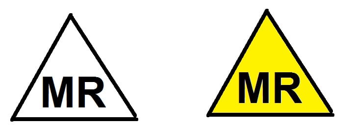
Examples:

  • Earplugs
  • Non-conductive Sheet
  • Plastic Slide Board

MR Conditional

This defines any object that is not completely MR Safe. This defines any object that is metallic, magnetic, and/or electrically conductive.
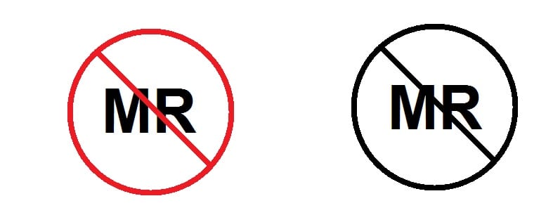
Examples:

  • Titanium joint replacement
  • MRI wheelchair
  • MRI fire extinguisher

MR Unsafe

This defines any object that has been tested and shown to be a threat to a patient or any object that has not been tested.

Example:

  • Ferrous aneurysm clip
  • Firearm
  • Electrically conductive clothing

American Board of MR Safety (ABMRS) Roles

MR Safety Officer (MRSO)

This individual is in charge of carrying out the policies approved by the MRMD. They will have a large amount of information on risk assessment and will help with providing information to the attending radiologist so that a decision with regards to scanning a patient can be made.

MR Medical Director (MRMD)

This individual is a physician who is in charge of the MR safety program at a site. They will appoint an MRSO and be a resource to attending radiologists with regards to threat evaluation and help with a benefit-risk assessment. They must review safety polices annually.

MR Safety Expert (MRSE)

This individual is the final resort when trying to investigate and/or evaluate the threats associated with an implanted device/ devices. Typically a physicist, they will adopt a scientific approach while uncovering threats to facilitate safe scanning. They will have an in-depth knowledge of physics. This is strictly a consultancy role/job.
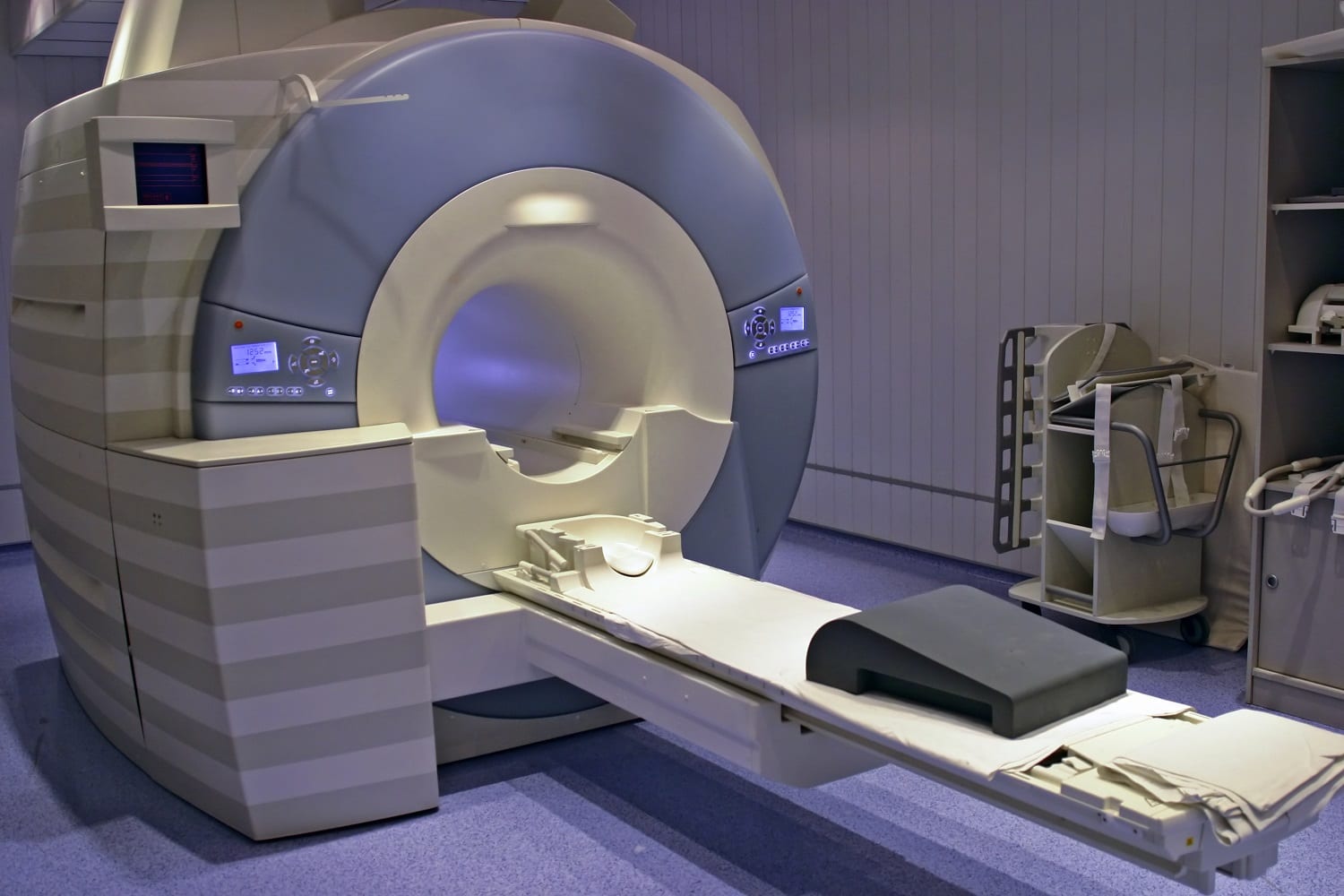
IEC Mode Of Operation

Whole Body RF Coil

The RF coil that is built into the MRI unit. It will transmit RF energy to our patient and has the capability to receive patient signal.

Local RF Coil

This RF coil will surround the body part of interest. It will transmit RF and receive signal from our patient.

Normal Operation Mode

Should Not Cause Physiological Stress To Our Patient

Whole Body SAR

Body= 2 W/kg

Head= 3.2 W/kg


Local SAR

Head and Torso= 10 W/kg

Extremities= 20 W/kg

First Level Controlled Operation Mode

May Cause Physiological Stress To Our Patient. Requires Medical Supervision.

Whole Body SAR

Body= 4 W/kg

Head= 3.2 W/kg


Local SAR

Head and Torso= 20 W/kg

Extremities= 40 W/kg

Second Level Controlled Operation Mode

A Level Beyond First Level Control Operation Mode

Whole Body SAR

Body= >4 W/kg

Head= >3.2 W/kg


Local SAR

Head and Torso= >20 W/kg

Extremities= >40 W/kg

Get the #1 MR Safety Training Course

Stop worrying about if that implant will hurt your patient!

MRSO Prep-01
www.RITEAdvantage.com
5/5
en_USEnglish (United States)

What the heck does this look like to you!?

These are not recommendations of MRI Buzz or any collaborators. Please check with your radiologist prior to delivering dose.