
Neurological Pathology
Stroke
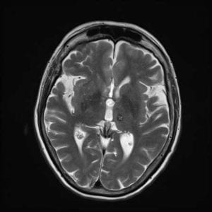
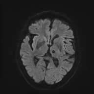
Acute Stroke
Middle Cerebral Infarct
Bleed
Bleed
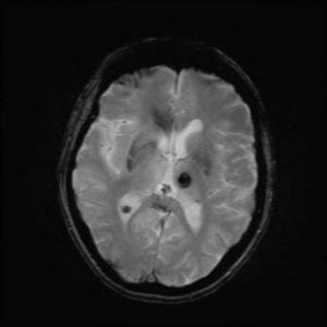
Gradient Echo T2*
T1 Weighting
T2 Weighting
Diffusion Weighting Tracer Image
Meningioma

Cyst
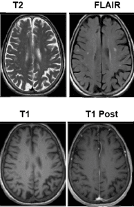
Multiple Sclerosis
Chiari Malformation
Abdominal Pathology
Polycystic Disease
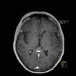
Enhancing Mass
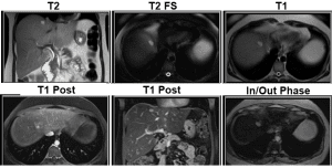
Hemangioma
The Bac and Christos Vault
Has Been Unlocked
Check More From:
Christos Tsiotsios
Head and Neuro
Stages of Brain Hemorrhage in MRI.
Cinematic rendering MRI and MRA of the brain using @siemens.healthineers Turbo Suite.
With Turbo Suite you accelerate the complete exam, covering all contrasts, all orientations, static and dynamic imaging, leading to up to 50% shorter exams.
Vestibular Schwannoma. The ice cream cone sign refers to the appearance of a medium-sized (1.5 to 3.0 cm) vestibular schwannoma. The intracanalicular component represents the cone and the cerebellopontine angle (CPA) (cisternal) component representing the ice cream ball.
Severe traumatic spinal cord injury with complete anterior dislocation of C6 over C7 vertebra (DOI: 10.7860/JCDR/2014/9471.5181)
High resolution T2*-Weighted MR imaging in a 7T, Siemens MAGNETOM Terra
High resolution MR Imaging of the Cervical Spine and MR Angiography (arteriography and venography) of the Neck in a Patient with Meningioma.
Image dataset acquired at 1.5T, GE SIGNA HDxt.
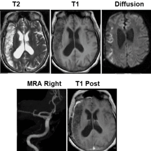
Giant Aneurysm of the Right Middle Cerebral Artery
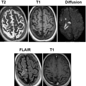
Patient with Brain Infarction.
Diffusion weighted imaging (DWI) is a commonly performed MRI sequence for evaluation of acute ischemic stroke, and is sensitive in the detection of small and early infarcts. Conventional MRI sequences (eg FLAIR) may not demonstrate an infarct for 6 hours, and small infarcts may be hard to estimate on CT for days.
High resolution Magnetic Resonance Neurography (MRN) of the Brachial Plexus, based on Coronal and Axial 2D STIR Acquisitions and Axial 2D T2-W Acquisition. Newer techniques like 3D STIR, 3D DIR or 3D NerveVIEW were not available (Image dataset acquired in an old 1.5T scanner).
The brachial plexus is a complex network of nerves formed by the anterior rami of the lower four cervical nerves (C5, C6, C7, C8) and first thoracic nerve (T1), which supplies motor and sensory innervation to the upper limb and pectoral girdle.
MRN is a non-invasive imaging technique for the dedicated assessment of peripheral nerves. Because of its excellent soft-tissue contrast, MRN offers anatomical information that is not obtainable with other modalities.
Read our EPOS and learn more about the MR Neurography of the brachial plexus: http://dx.doi.org/10.26044/ecr2019/C-2391
Sagittal T2-weighted MR Image demonstrates Corpus Callosum Lipoma and Huge Subcutaneous Lipoma in an Adult. Irregular anterior cerebral artery can also be seen.
DOI: https://dx.doi.org/10.4103%2F0976-3147.127932
MR Neurography of the Brachial Plexus in a Patient that referred to MRI Post Trauma of the Head-Neck region. Image courtesy of @philips.
Pathologically- proven Schwannoma of Brachial Plexus in a 40-year-old Patient with Right Arm Dysesthesia.
(A) 3D SPACE STIR volume rending (VR) image of the brachial plexus revealed a round mass (arrow) in the upper trunk.
(B) The tumor showed inhomogeneous contrast enhancement on the venous phase coronal VIBE image (arrows head).
(C) 3D-Synchroview demonstrates the relationship of the mass to the surrounding vessels, and the subclavian artery was compressed and displaced (arrow).
Full-text PDF: https://www.medscimonit.com/abstract/index/idArt/919358
The first presumptive case of COVID-19–associated Acute Necrotizing Hemorrhagic Encephalopathy, a rare Encephalopathy that has been associated with other viral infections but has yet to be demonstrated as a result of COVID-19 infection (Poyiadji N. et al., 2020). DOI: https://doi.org/10.1148/radiol.2020201187
T2-Weighted Imaging of the Hippocampus: 3T vs 7T
Very High Spatial Resolution Axial T2-weighted Image of the Brain with 0.1mm x 0.1mm In-Plane Resolution.
Image acquired at 3T, uMR 790, courtesy of United Imaging.
Giant Medial Sphenoid Wing Meningioma
3D NerveVIEW for Non-Invasive Assessment of the Brachial Plexus! Courtesy of @philips.
Diffusion Tensor Imaging (DTI) with 1mm isotropic resolution covering the Whole Brain. Image dataset acquired at 7T, courtesy of @siemens.healthineers.
Cinematic Rendering of a Magnetic Resonance (MR) Image computed from Diffusion Tensor Imaging (DTI) of the brain. Image dataset acquired at 7T, courtesy of @siemens.healthineers.
High resolution, Dedicated MR Imaging of the Orbits in a Patient with Orbital Melanoma. Image dataset acquired at 3T, GE SIGNA Architect.
Primary or secondary orbital melanomas are extremely rare tumors; they represent less than 1% of primary orbital neoplasms. Over 90% of primary orbital melanomas arise from melanocytes.
High-Resolution Anatomical MR Imaging of the Brain in a Patient with Glioblastoma Multiforme (GBM).
Figure shows a volumetric FLAIR CUBE acquisition, two 2D T2-weighted PROPELLER acquisitions in coronal and axial planes and a post-contrast volumetric T1-weighted CUBE acquisition. PROPELLER technique was used in the 2D T2-w sequences in order to reduce motion-related artifacts.
Image dataset acquired at 3T, GE Architect, using the 48ch head coil.
Brain MRI Slice-Positioning
The Brain Vessels of a Volunteer are imaged using a 3T (left) and 9.4T (right) MRI scanner.
Credit: Rolf Pohmann/Max-Planck-Institute for Biological Cybernetics
DOI: 10.1038/d41586-018-07182-7
Very High Resolution, Small FOV MR Imaging of the Orbits in a Patient with Cavernous Venous Malformation. Image dataset acquired on a 12-year-old GE Signa HDxt 1.5T.
The cavernous venous malformation of the orbit, previously called cavernous hemangioma, is the most common primary orbital lesion of adults. Cavernous venous malformation occurs more often in women and typically presents in the fourth and fifth decades of life. It is a benign vascular malformation characterized by a well-defined capsule and numerous large vascular channels.
High Resolution FLAIR image from a Multiple Sclerosis (MS) Patient, acquired at 7T. The white matter lesion shows the central vein sign.
Image courtesy of @siemens.healthineers.
Various representations of the Human Head/Brain, Magnetic Resonance Tomography with a field strength of 7 Tesla (Post-Processing with”Cinematic-Rendering").
Image courtesy of @siemens.healthineers.
Post-Contrasted Sagittal T1-W Imaging of a Patient with a large vein of Galen Aneurysmal Malformation (red arrow).
DOI: 10.7759/cureus.2180
Hemispherectomy is a very rare neurosurgical procedure in which a cerebral hemisphere (half of the brain) is removed, disconnected, or disabled. This procedure is used to treat a variety of seizure disorders where the source of the epilepsy is localized to a broad area of a single hemisphere of the brain, notably Rasmussen's encephalitis.
About one in three patients with epilepsy will continue to have persistent seizures despite epileptic drug therapy. Hemispherectomy is reserved for the most extreme cases of this one-third in which the individual’s seizures are irresponsive to medications or other less invasive surgeries and significantly impair functioning or put the patient at risk of further complications. The procedure successfully cures seizures in about 85–90% of patients.
Image courtesy of Dorit Kliemann et al. (DOI: https://doi.org/10.1016/j.celrep.2019.10.067)
Sagittal T1 Brain MRI with Contrast showing Huge Extra-Axial Dural Base Enhance Lesion with Dural Tail and Compression on Adjacent Structures.
DOI:10.1136/bcr-2017-220833
Thick MIP of a 3D SPACE STIR sequence in a Patient with Charcot Marie Tooth Disease (CMT).
Courtesy of @siemens.healthineers.
Neuroradiological findings in Adult Cranially-Conjoined Twins (Jansen O. et al).
DOI: https://doi.org/10.3171/jns.1998.89.4.0635
T1-Weighted Magnetic Resonance Image (MRI) of a Middle-Aged woman with Progressive Headaches, Aphasia, and Right-sided Hemiparesis. A large intracerebral mass with a significant amount of surrounding edema is depicted. The lesion is a giant internal carotid artery aneurysm.
https://emedicine.medscape.com/article/337027-overview
Coronal 3D STIR Oblique MIP. Image courtesy of @philips.
Holocord Syrinx associated with Haemangioblastoma.
DOI: http://dx.doi.org/10.1136/practneurol-2015-001251
Magnetic Resonance Imaging shows a large heterogeneous soft tissue lesion with flow void seen at the periorbital and intraconal parts of the left orbit in keeping with arteriovenous malformation.
It is causing displacement of the left globe anterolaterally outside the bony orbit. This arteriovenous malformation extends medially to involve the nasal bridge and superiorly to involve the scalp.
DOI: https://doi.org/10.1016/j.asjsur.2013.09.011
Multi-Cystic Encephalomalacia in a 2 month-old Infant.
DOI: http://dx.doi.org/10.1594/ecr2018/C-3167
A 50-year-old Male who has been suffering headaches and strange smells has finally discovered the cause of his woes: A 1 cm (0.4 inch) Tapeworm in his Brain.
The parasite is thought to have been burrowing through his grey matter for more than four years, moving 5 cm (2 inches) from the right side to the left.
Aneurysmal Bone Cyst of Skull Base.
http://www.rsroc.org.tw/db/Jrs/article/V35/N1/350106.pdf
MR Imaging of the Spine on a Patient with Scoliosis.
Scoliosis is a sideways curvature of the spine that occurs most often during the growth spurt just before puberty. While scoliosis can be caused by conditions such as cerebral palsy and muscular dystrophy, the cause of most scoliosis is unknown. About 3% of adolescents have scoliosis. Most cases of scoliosis are mild, but some spine deformities continue to get more severe as children grow.
Agenesis of the Corpus callosum (ACC).
It is a rare birth defect (congenital disorder) in which there is a complete or partial absence of the corpus callosum. It occurs when the corpus callosum fails to develop normally, typically during pregnancy. The fibers that would otherwise form the corpus callosum become longitudinally oriented along the ipsilateral ventricular wall and form structures called Probst bundles.
Sagittal T2-weighted MR image of the Brain. Variant Dandy–Walker Syndrome (DWS) with Dysplasia of the Pons and Cerebellum in an 8-year old.
DWS is a rare group of congenital human brain malformations. There are three subtypes which affect multiple organs to varying degrees, but the fundamental abnormalities involve the cerebellum which controls muscle coordination. The adjacent third and fourth ventricles are often affected, which can alter the flow of cerebrospinal fluid, increase intracranial pressure and lead to multiple other brain function problems.
CSF-filled Brain Lesion detected by Sagittal 3D BrainVIEW FLAIR.
Courtesy of @philips
High-Resolution Vascular Imaging (based on SWI) of a Patient with Brain Tumor showing detailed visualization of the vasculature within and around the pathologically affected tissue.
Image dataset acquired at 7T, courtesy of @siemens.healthineers.
High-Resolution Anatomical and Functional MR Imaging of the Cervical Spine and Brachial Plexus in a Patient with Malignant Peripheral Nerve Sheath Tumor (MPNST).
MPNST is a kind of highly aggressive neoplasm originated from Schwann cells. As the name implies, MPNST develops in the peripheral nerves and scarcely affects the cranial nerves.
MRI is a valuable, non-invasive modality for the diagnosis and follow-up of MPNST.
Diffusion Tensor Imaging (DTI) of the Lumbosacral Plexus, obtained with Read-out Segmented EPI technique, in order to minimize image distortions.
DTI can show if a tumor deviates, infiltrates or distructs the fiber bundles, offering functional information. Image dataset acquired at 3 Tesla, Courtesy of Bac Nguyen.
Patient with Giant Trabecular-type Juvenile Ossifying Fibroma of the Maxilla. MRI scan of the mass did not show any intra-cranial extension.
https://www.elsevier.es/es-revista-revista-espanola-cirugia-oral-maxilofacial-300-articulo-giant-trabecular-type-juvenile-ossifying-S1130055816300466
DOI: 10.1016/j.maxilo.2016.10.003
Parameningeal Rhabdomyosarcoma.
Coronal T2-W (a) and axial T1-W (b) images show a 12 × 10 cm lobulated, heterogeneously enhancing (c) right facial mass extending from the base of skull to the inferior margin of the mandible.
Rhabdomyosarcoma is a malignant tumor with skeletal muscle cell morphology. Rhabdomyosarcomas are the most common soft tissue tumor in children and account for 5-8% of childhood cancers, and 19% of all pediatric soft tissue sarcomas (du Toit J, Wieselthaler N, 2015).
Giant Intracranial Hydatid Cyst in an 11-Year Old Boy.
(Waziri T.M. et al. 2017).
Sagittal T2-W MR Imaging shows Fracture Dislocation at T4-T5 level with Spondylolisthesis of T4 over T5, and Contusion of Cord.
Umesh C Parashari et al. 2011.
T2-weighted axial MRI of a Giant Meningioma - Pediatric patient.
Giant Basilar Artery Aneurysm causing Bulbar Palsy and Hydrocephalus (BMJ 2018)
Sagittal T1-W W/O Contrast shows a Giant Pituitary Adenoma with extension to the Nasopharynx, Sphenoid, and Ethmoid sinuses.
Axial T2 (A), Axial T1 Pre (B) and Post-Contrast (C), and Sagittal T1 Post-Contrast (D) MRI Images demonstrate large well-defined Multi-loculated Complex Neck Mass (Congenital Cervical Teratoma) with Significant Mass Effect.
The solid component (arrows) shows intermediate T2 and T1 signal intensity with avid heterogeneous enhancement while the cystic component (asterisks) shows high T2 and low T1 signal intensity with no enhancement. Oesophagus (arrowhead) and airway (dashed arrow) are displaced (Alharbi S. et al. 2018).
Composite Image of the Brain from a Healthy Young Adult. The Right Hemisphere is a 3D reconstruction from Structural 3T Magnetic Resonance Imaging (MRI) Data (pink), while in the Left Hemisphere, Nerve fibres have been visualised using Diffusion Imaging Tractography (multi-coloured).
Diffusion tensor imaging (DTI) is a specialised type of magnetic resonance imaging (MRI) scan which measures water diffusion in many directions in order to reconstruct the orientation of bundles of axons. Tractography is used to indirectly model these bundles of axons (nerve fibres), which transmit information between cortical regions at the brain's surface.
Credit: Gabriel González-Escamilla
Patient with Arteriovenous malformation (AVM).
AVM is an abnormal connection between arteries and veins, bypassing the capillary system.
SWI Contrast in MRI.
SWI is a new type of contrast in MRI which exploits the susceptibility differences between tissues. As a result SWI detects substances with different susceptibilities than their neighboring tissues such as deoxygenated blood, products of blood decomposition and microscopic iron deposits much better than conventional "hemo" MR techniques.
Among other things, the method allows for highly sensitive proof of cerebral hemorrhage and high resolution display of venous cerebral vessels.
Malignant Peripheral Nerve Sheath Tumor in Neurofibromatosis Type 1 (Rossiter J. et al., 2014)
NerveVIEW is a high resolution T2-w TSE acquisition with reduced intra-lumen signal of the veins, which improves the visualization of the brachial and lumbosacral plexus.
In addition, the 3D isotropic imaging method allows for reformats in any plane (including oblique) without loss of resolution.
Multiparametric MR Imaging of Brain's Meningioma.
Based on high-resolution morphological (axial T2 PROPELLER and IR-prepared 3D T1 GRE pre and post contrast are shown here) and functional (DWI b1000 and DSC PWI are shown here, also DCE PWI was acquired) acquisitions. The high rCBV and rCBF of the lesion should be noted.
Figure illustrates the effect of different echo times (TE) on image signal-to-noise ratio (SNR). Images were acquired using a pseudo patient head phantom.
Images courtesy of Christos Tsiotsios.
Intracranial 4D flow MRA. Non-contrast enhanced blood flow visualisation and quantification in a large Arteriovenous Malformation (AVM).
HyperBand DTI Tractography fused with HyperSense 3D TOF (Volume rendering).
Two Most Common Cardiac Tumors.
(A-C) Cardiac Angiosarcoma and (D-F) Cardiac Lymphoma (Colin G. et al. 2015).
Primary cardiac malignancies are extremely rare.
Neuroimaging at 7T. FLAIR (left) and 3D SWI (right) exhibiting Multiple Sclerosis (MS) lesions in the White Matter, but also in the Cortex.
Sagittal T2-weighted MRI shows Uterine leiomyomatosis with the whole uterus transformed to numerous small fibroids (white arrows) (Duc NM, Huy HQ, 2018).
Patient with Severe Thoracic Kyphosis.
MRI of the cervical and thoracic spine (left: T1-w, right: STIR) shows pronounced osteoporotic collapse of T12 with dislocation of the spinal cord.
CT and zero TE (ZTE) MR images acquired from a Molar with Caries Lesions. Some residual bone attached to the teeth is visible.
Occlusal demineralized lesion (arrows A,B), Halo effect around the pulp (arrow C), the fissures visible with mCT inside the dentine (arrow D), demineralized lesions (arrow E).
(Weiger M. et al. High-resolution ZTE imaging of human teeth. NMR Biomed 2012;25(10):1148)
Teeth in the brain’ – A Case of Giant Intracranial Mature Cystic Teratoma (O'Grady J. et al., 2012)
Axial T1- and T2-weighted MR Images demonstrate Bilateral Cleft communicating with the Subarachnoid spaces and lined by Heterotopic Gray Matter. The characteristic batwing appearance is noted (Omidiji et al. 2018).
Axial T2-weighted MR image of the Orbits show a Large Extraconal Mass pushing the globe inferotemporally and it breaches the cribriform plate to extend intracranially into both frontal lobes (Ainal Adlin N et al., Journal of Surgical Academia 2016; 6(1): 59-61).
DTI (Fiber-tracking Map/Tractography) of the Sacral Plexus (left) and Sciatic Nerve (right).
DTI depicts the anisotropic diffusion and can provide useful structural information about the nerves. Image dataset acquired at 3.0 Tesla. Images courtesy of Bac Nguyen.
Patient with Arteriovenous Malformation (AVM).
Anatomical and functional sequences were used here for the pre-surgical mapping and assessment of the lesion! Image post-processing via Nordic Neuro Lab (NNL). Courtesy: S. Magnetic World
Modified 3D FSE (CUBE/SPACE/VISTA) Imaging.
It can replace slice-by-slice, plane-after-plane, 2D acquisitions with one single 3D volume scan. The operator can easily reformat sub-millimeter, isotropic 3D volume data into any plane without gaps, and with (approximately) the same resolution as the native plane. Moreover, modified 3D FSE sequences offer similar contrast as 2D acquisitions.
Clinical applications: • Neuro• MSK • Body imaging
Image courtesy of GE healthcare.
CUBE DIR for Imaging the Brachial Plexus.
3D DIR is a volumetric acquisition that provides both fat and CSF suppression, which leads to an improved nerve-to-background tissue contrast and enhanced ability to visualize the brachial plexus.
Image courtesy of GE Healthcare.
MRE Wave and Elastogram Images compared in a Normal Subject (upper row) and a Patient with Hepatic Fibrosis (lower row)
MRE allows estimation of the mechanical properties of tissue in response to compression or vibration. Available as a commercial product from at least three MR vendors, its current principal application is for staging hepatic fibrosis. (Li et al 2015).
MR Imaging of the Brain on a Patient with Astrocytoma.
Standard post-contrast T1-w SE shows uptake of the contrast media, the AUC map (extracted from DCE Permeability Imaging) presents neoangiogenesis, while DTI Tractography highlights that the tumour distracts the fiber bundles.
Cartesian 2D T2-w FLAIR (A) and non-Cartesian (PROPELLER) 2D T2-w FLAIR (B). Cartesian acquisition suffers from severe motion artifacts, while non-Cartesian acquisition easily highlights the cerebellum tumor, offering diagnostic imaging.
Images courtesy of Christos Tsiotsios.
Cinematic – Internal Acoustic Canal (IAC) at Siemens Skyra 3 Tesla. Courtesy: Bac Nguyen, S. Magnetic World.
MRI of Fractured and Dislocated Neck Vertebra that is compressing the Spinal Cord.
Arnold-Chiari Malformation Type III With Meningoencephalocele (Jeong DH et al. 2014)
Volumetric T2-W and T1-W (+C) on a patient with Glioblastoma Multiforme (GBM). Image dataset acquired at 1.5T.
Whole body MR Neurography
(Yamashita T, et al. N Engl J Med. 2009)
Blood oxygenation level dependent (BOLD) MR Imaging.
This is the standard technique used to generate images in functional Magnetic Resonance Imaging (fMRI) studies, and relies on regional differences in cerebral blood flow to delineate regional activity.
MR Lymphangiography at 1.5 Tesla.
Courtesy: Bac Nguyen, S. Magnetic World.
Comparing DWI TSE & SS-EPI DWI for Spinal Pathology MRIs.
DWI TSE allows diffusion imaging with excellent signal-to-noise ratio and sharpness with reduced geometric distortion compared to SS-EPI DWI, especially in challenging anatomies such as spine.
Image fusion: fMRI, DTI and anatomical 3D T1-W.
Philips Ingenia 1.5T. Image courtesy of @philips
High Resolution Meningioma MR Imaging.
Based on anatomical and functional acquisitions.
DWI PROPELLER makes feasible the DW Imaging of inhomogeneous areas, such as IAC, without distortions. Thin MIP of the volumetric T1 acquisition can be used for the (time-effective) evaluation of vessels proximity.
Images courtesy of Christos Tsiotsios.
High Diffusion and Spatial Resolution Diffusion Tensor Imaging (DTI) of the brain (1.5mm isotropic voxel size, 64 diffusion directions).
Colour-coded orientation map fused on an anatomical 3D T1-w MR image (A, B, C) and whole brain fiber-tracking map (D).
Slice acceleration (SMS, Multi-Band) can be used to decrease the scan time and/or achieve higher spatial/diffusion resolution. High diffusion resolution is necessary for advanced diffusion models (e.g. Kurtosis) and for advanced post-processing procedures (e.g. constrained spherical deconvolution, which addresses the crossing fibers issue).
Image dataset acquired at 3 Tesla. Courtesy of Bac Nguyen.
MR Imaging of the Brain in a Patient with Glioblastoma Multiforme (GBM).
High resolution anatomical sequences (volumetric T2, volumetric FLAIR, volumetric T1), 3D SWAN (for hemorrhage and vasculature assessment) and functional-quantitative sequences (DWI and PWI) was used.
GBM (also called glioblastoma) is a fast-growing glioma that develops from star-shaped glial cells (astrocytes and oligodendrocytes) that support the health of the nerve cells within the brain. These are the most invasive type of glial tumors, rapidly growing and commonly spreading into nearby brain tissue.
In adults, GBM occurs most often in the cerebral hemispheres, especially in the frontal and temporal lobes of the brain.
MR diffusion-weighted imaging (DWI) in monitoring the response to treatment in Cervical Cancers.
DWI shows that there is an increase in tumor's diffusivity (the ADC value before radiotherapy was 603 μm2/sec, and the ADC value after radiotherapy is 917 μm2/sec) and a decrease in tumor's volume after the radiotherapy. These changes in tumor's microarchitecture are not initially seen using conventional anatomical imaging.
Multiple Liver Metastases in a Patient with Breast Cancer. Images courtesy of Christos Tsiotsios.
Figure shows a patient with prostate cancer (adenocarcinoma), with low signal intensity on axial T2-w, high signal on high b-value DWI, low signal on corresponding ADC map (ADC value of 598 μm2/sec) and fast wash-in on DCE PWI. Patient will undergo U/S-guided brachytherapy.
The recommended use of MRI in prostate cancer detection, localization, staging and characterization consists of multiparametric MRI (mpMRI).
MpMRI refers to the use of anatomical imaging (T2-w and T1-w) in combination with functional and quantitative imaging techniques (DWI, DCE PWI and optional MRSI)
Images courtesy of Christos Tsiotsios.
Another Image highlighting - Signal, Contrast and Resolution Information in K-space.
Abdomen/Pelvis/Body
High resolution T2-weighted and DWI MR images of the Prostate and Penis in a Patient with Prostate Cancer, metastasizing to the Corpus Cavernosum.
Despite the rich vascularisation and complex circulation, metastasis to the penis is rare, and most commonly arises from the prostate and the bladder.
The most common symptoms are penile induration, swelling and palpable nodular lesions on penis. These patients have a poor prognosis receiving various treatment modalities.
Image dataset acquired at 3T, GE SIGNA Architect.
High Resolution, Multiparametric (anatomical, dynamic and functional) Gadoxetate Disodium-Enhanced MR Imaging of the Liver in a Patient with Hepatocellular Carcinoma (HCC).
Image dataset acquired at 1.5T, GE SIGNA HDxt. The total examination time was 20min.
Figure illustrates the usefulness of Whole Body MRI (WB-MRI), and shows a Patient with Malignant Melanoma and Widespread Metastases including the Brain.
WB MR Imaging shows Multiple Lesions outside the FOV of CT thorax and abdomen:
(a) A Maximum Intensity Projection (MIP) image of DWI shows the disease distribution in a valuable overview of the entire body.
(b) Coronal STIR, and
(c) Coronal T1-Wsequences, respectively, show further anatomical details of metastatic lesions (Mosavi F. et al. 2013).
Patient with elevated PSA and a very suspicious area in the transition zone with low signal on T2-w, diffusion restriction and high perfusion and permeability, as indicated by the quantitative analysis of DCE-MRI data.
The role of Dynamic Contrast-Enhanced Magnetic Resonance Imaging (DCE-MRI) for tumour diagnosis and assessment in the current version of PI-RADS (version 2) is minimized. DCE-MRI is only considered when DWI in the peripheral zone (PZ) is indeterminate.
Although, DCE Perfusion has shown potential in prostate cancer detection, localization and staging, assessment of tumour aggressiveness and assessment of treatment response.
Note that ktrans is the influx mass transfer rate of gadolinium (ktrans depends on blood flow, capillary surface area, and capillary permeability), and kep is the corresponding efflux rate of gadolinium from the extravascular-extracellular space (EES) back into plasma.
MR-Enterography (MRE) is a non-invasive technique for diagnosis of Small Bowel Disorders.
MRE is most commonly used to evaluate Patients with Crohn’s Disease where it is used for assessment of the primary disease and any complications.
Very High Resolution T1 FLASH FatSat, Matrix 512, GRAPPA 2.
Image courtesy of @siemens.healthineers.
Whole body MRI of Neurofibromatosis type I (NF-1).
Giant Warthin’s Tumour of the Parotid Gland.
Warthin’s tumour was first adequately described in 1929 by an American pathologist, Aldred Scott Warthin. It is synonymous with papillary cystadenoma lymphomatosum (PCL), cystadenolymphoma or adenolymphoma.
Giant salivary gland tumours are usually pleomorphic adenomas. The average size of Warthin’s tumour is reported to be about 2–4 cm.
DOI:10.1007/s12070-019-01675-1
The Horseshoe Kidney - Most common type of Renal Fusion Anomaly.
It consists of two distinct functioning kidneys on each side of the midline, connected at the lower poles (or rarely at the upper poles) by an isthmus of functioning renal parenchyma or fibrous tissue that crosses the midline of the body.
Magnetic Resonance Enterography (MRE). Figure shows a T2-Weighted Single-Shot Fast Spin Echo/SS-FSE (left) and a Volume-interpolated 3D T1-w GRE DIXON (right).
Oral contrast agents must be used in MRE.
Oral contrast agents used for MRE can be categorized as positive, negative, and biphasic (figure) contrast agents.
The most commonly used MRE contrast agents are biphasic, exhibiting high signal intensity on T2-w imaging (allowing for detection of wall thickening, endoluminal abnormalities, and transmural ulcers) and low signal intensity on T1-w imaging (enhancing detection of mucosal enhancement and hypervascular endoluminal masses).
Image courtesy of Bac Nguyen, S.Magnetic World.
Situs inversus, short form of the Latin “situs inversus viscerum”, is a term used to describe the inverted position of chest and abdominal organs.
It is called Situs inversus totalis when there is a total transposition of abdominal and thoracic viscera (mirror image of internal organs normal positioning).
24-Year Old Male with Large Hemorrhagic Abdominoscrotal Hydrocele.
TSE T2-Weighted Sagittal Plane. The caudal-cranial mass (about 23 cm) extends in the abdomen through the right inguinal canal to the top of L5. Since the mass can not expand in the right inguinal ring, on this view the lesion appears "hourglass" shaped. Posteriorly to the mass it is possible to recognize the bladder. Just below the mass on the right side, there is a hydrocele with fluid signal intensity.
DOI: 10.3941/jrcr.v3i6.132
3D MRCP showing Significant Dilation of Bile Ducts, right (blue) left (red) and common bile duct (purple) (Ovungu JM et al. 2017).
Comparison of different sequences and flip angles used for Excretory MR Urography.
(a) On a Coronal MIP image from Excretory MR Urographic data obtained with a 3D interpolated fat-suppressed gradient-echo sequence (VIBE/THRIVE/LAVA) during breath holding, soft-tissue suppression is minimized owing to the use of a relatively low flip angle of 12°.
(b) Coronal MIP image from Excretory MR Urographic data obtained with a 3D gradient-echo MR angiographic sequence shows improved background tissue suppression due in part to the use of a higher flip angle of 40° (Leyendecker et al 2008).
You can find more info about MRU here: https://www.myradiologyedu.com/post/mr-urography-patient-preparation-technique-and-pulse-sequences
Coronal Contrast-Enhanced T1-W TSE mDIXON (water only) on a Pediatric Patient with Head/Neck Tumor.
Image courtesy of @philips.
High Resolution MRCP T2 SPACE at Siemens Magnetom Vida, 3T.
Courtesy: Siemens Healthineers, S.Magnetic World
A Bicornuate Uterus or Bicornate Uterus, is a type of Mullerian anomaly in the Human Uterus, where there is a deep indentation at the Fundus (top) of the Uterus.
Diagnosis of bicornuate uterus typically involves imaging of the uterus with 2D or 3D ultrasound, hysterosalpingography, or MRI.
(Carrascosa P. et al. 2014)
Axial T1-Weighted Sequence shows a left Multiloculated, Heterogeneous Cystic Mass that contains Cyst-like areas of variable size extending into the Right Orbit and Temporal Lobe (Mature Teratoma) (Nejat Isik et al. 2012)
Magnetic resonance cholangiopancreatography (MRCP) Image.
MRCP is a medical imaging technique that uses magnetic resonance imaging to visualize the biliary and pancreatic ducts in a non-invasive manner.
MRCP makes use of heavily T2-weighted MRI pulse sequences. These sequences show high signal in static or slow moving fluids within the gallbladder, biliary ducts and pancreatic duct, with low signal of surrounding tissue.
In the diagnosis of pancreatic disorders, MRCP is a much less invasive investigation when compared to endoscopic retrograde cholangiopancreatography (ERCP).
Although both techniques can image the ductal system in detail, MRCP also allows imaging of the surrounding parenchyma.
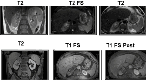
Autosomal Dominant Polycystic Kidney Disease (ADPKD).
In ADPKD the kidneys and liver can be extremely enlarged due to cyst formation, with a single kidney volume increasing up to 6,000 mL and a liver volume up to 14,000 mL.
MIP Contrast-Enhanced Breast MR image which illustrates a Large Left Breast Malignant Phyllodes Tumor.
Very high resolution T2-Weighted imaging of the Prostate Gland. Image dataset acquired at 3T, Siemens Vida.
Gadoxetate Disodium–Enhanced (Primovist/Eovist) MR Imaging of Small Liver Metastasis.
The combination of hepato-specific contrast agent with anatomical and functional imaging techniques can greatly increase the sensitivity and specificity of the examination.
Images courtesy of Christos Tsiotsios.
Figure illustrates the three possible enhancement kinetic curves in Breast MR Imaging (can also applied to other organs such as prostate), after the administration of Gadolinium-based contrast agent (GBCA).
a) Curve type 1: progressive enhancement pattern
• Typically shows a continuous increase in signal intensity throughout time.
• Usually considered benign with only a small proportion of (5-10%) of malignant lesions having this pattern.
b) Curve type 2: plateau pattern
• Initial uptake (slow or fast) followed by plateau phase towards the delayed phases of the dynamic study.
• Suspected for malignancy.
c) Curve type 3: washout pattern
• Has a relatively fast uptake (wash-in) and shows reduction in enhancement (wash-out) towards the delayed phases of the dynamic study.
• Considered strongly suggestive of malignancy.
Images courtesy of Christos Tsiotsios.
High-resolution, high-quality, multiparametric (anatomical and functional) Prostate MR Imaging in a Patient with Elevated PSA.
Orthogonal high resolution T2-w FSE, high b-value DWI (acquired not calculated), ADC map and postcontrast fat-suppressed 3D T1-W GRE are shown here.
Images courtesy of Christos Tsiotsios.
High-resolution, Multiparametric (morphological and functional) MR Imaging of the Pelvis.
Figure illustrates a pelvic lymph node metastasis (red circle), characterized by hypercellularity (low ADC) and Gd uptake.
High Resolution, Multiparametric Magnetic Resonance Enterography (MRE) in a Patient with Crohn's disease (CD).
Crohn's disease is most commonly found in the terminal ileum (red circle) and colonic region. MRE has become a useful modality for assessing small bowel activity.
Click here, read our electronic poster, and learn everything you need to know about MR Enterography: http://dx.doi.org/10.26044/ecr2020/C-02140
Figure shows the same 2D T2-w FS FSE acquisition.
3 NEX and respiratory triggering was used in the image to the left, while 1 NEX and breath holding technique was used in the image to the right. Note that the metastasis can clearly be seen only in the image to the left.
The number of excitations (NEX or NSA or averages) is used to represent the number of times each line of k-space data is acquired and is primarily used to improve the signal-to-noise ratio (SNR).
However, because the contrast-to-noise ratio (CNR) is controlled by the same factors that affect SNR, keep always in mind that NEX also affects the image CNR, as shown by this figure.
Whole-body Magnetic Resonance Imaging (WB-MRI).
Whole-body magnetic resonance imaging (WB-MRI) has been available for some time, but only recently it has become an efficient method for total body screening from head to toes.
In clinical practice, WB-MRI is increasingly being used in the diagnosis of a variety of diseases, including lymphomas, multiple myeloma, systemic musculoskeletal diseases, and also systemic diseases like diabetes and atherosclerosis. Furthermore, WB-MRI is very useful in children with malignant diseases (and other pathologies) due to the use of non ionising radiation.
Improvements of the software and hardware, multiplanar imaging capabilities, high contrast resolution, anatomical and functional imaging make MRI a great alternative for whole body screening within clinically acceptable acquisition times.
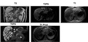
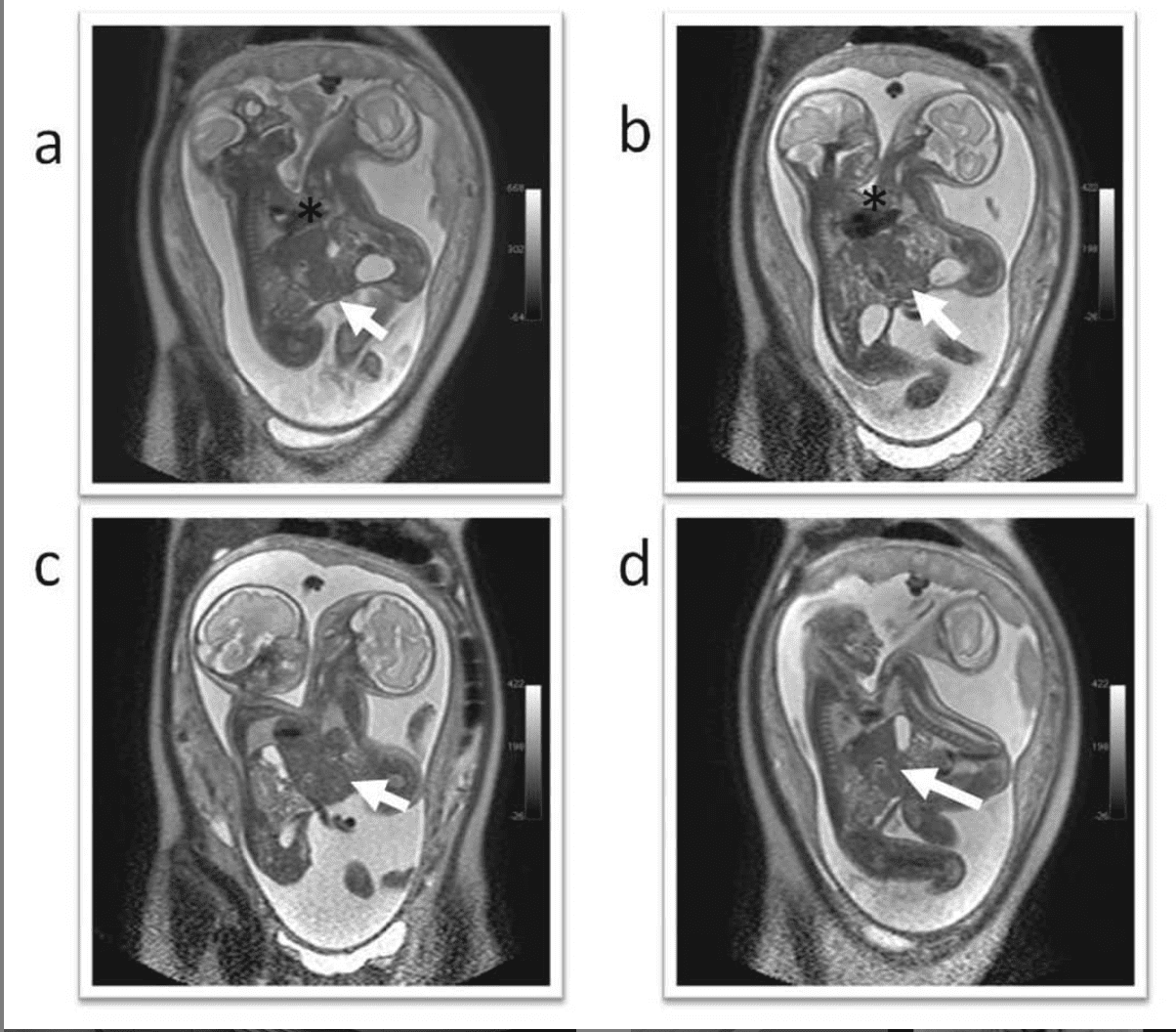
A 9-month-old Female Toddler with Hepatomegaly and Numerous Infantile Hepatic Hemangiomas.
(a) Color Doppler image through the left lobe of the liver shows multiple predominantly hypoechoic circumscribed masses with adjacent increased vascularity.
(b,c) Axial T1-weighted and T2-weighted fast spin echo fat-saturated images reveal numerous circumscribed T1-weighted hypointense and T2-weighted hyperintense liver lesions.
(d,e) Axial arterial phase and coronal delayed phase T1-weighted three-dimensional spoiled gradient recalled fat-saturated post-contrast MR images show early peripheral enhancement of the lesions with complete central fill-in over time.
(Hegde S. et al 2013).
Very High Spatiotemporal Resolution, Multiparametric (anatomical and functional) MR Imaging of the Prostate Gland on a Patient with Elevated PSA. The examination was performed on a 10-year-old, GE Signa HDxt 1.5T scanner.
Comprehensive, Multiparametric (anatomical and functional) MR Imaging of the Gynecological Organs on a big patient with Non-Keratinizing Squamous Cell Carcinoma of the Cervix.
Multiparametric gynecological organs protocol consists of high resolution T2-w acquisitions (at least in two imaging planes), diffusion-weighted imaging (low, intermediate and high b values are needed) with corresponding ADC map, dynamic contrast-enhanced perfusion-weighted imaging (DCE PWI), and pre- and post-contrast T1-w acquisitions.
High resolution MR Imaging of the Cirrhotic Liver at 1.5 Tesla.
Cirrhosis is scarring of the liver caused by long-term liver damage. The scar tissue prevents the liver working properly. Cirrhosis can eventually lead to liver failure, where the liver stops working, which can be fatal.
Unfortunately, conventional MRI is insensitive in early cirrhosis, and its role is in screening cirrhotic livers for small HCCs.
LiverMultiScan (Perspectum Diagnostics patented technology) can assist in the detection of early liver disease and predict the outcomes in patients with chronic liver disease.
More info about Perspectum and LiverMultiScan here: https://perspectum-diagnostics.com/
Images courtesy of Christos Tsiotsios.
High resolution MR Imaging of the liver in a patient with moderate ability of breath-holding, illustrating multiple lesions within the liver, consistent with metastatic disease.
That is why standard 3D T1 FS was used instead of 3D T1 Dixon.
Images courtesy of Christos Tsiotsios.
Gadoxetate Disodium–Enhanced MR Imaging of the Liver.
Figure presents a metastasis in right lobe of liver (VII segment). Late arterial phase shows fast wash-in, due to metastasis' hypervascularity, hepatocyte phase shows no uptake in this metastatic lesion and high b value DWI shows diffusion restriction.
Gadoxetate disodium (Primovist/Eovist) is an hepatospecific paramagnetic gadolinium-based contrast agent (GBCA), used exclusively in liver MRI. Its main uses are in assessing focal liver lesions and evaluating biliary leakage.
Images courtesy of Christos Tsiotsios.
Multiparametric MR Imaging of the Rectum for the Evaluation of Treatment Response.
DWI is great for pathology detection, and residual disease can be easily highlighted. Images courtesy of Christos Tsiotsios.
MRI of the Prostate Gland: Multiparametric Prostate MRI
MRI of the Prostate Gland was first described in the mid-1980s with early publications describing the sub-structure visualization and characterization using T2-w imaging.
Even with the low spatial resolution attained with the medium-field magnets and body coil technology employed at the time, it was immediately evident that MR could provide a unique and novel approach to imaging diseases of the prostate.
The endorectal coil, also introduced in the mid-to-late 1980s, added significantly to the quality of prostate MRI by enhancing visualization of sub-glandular structures.
In the last decade, Multiparametric Prostate MRI (which essentially combines anatomical and functional data) has seen a major increase in interest in the detection and characterization of focal prostate cancer with exciting developments in diffusion-weighted imaging (DWI) and dynamic contrast-enhanced perfusion-weighted imaging (DCE PWI), techniques that show promise in allowing for better assessment of prostate cancer aggressiveness through correlation with low, intermediate, and high Gleason scores.
Figure shows navigated Turbo LAVA, which provides free breathing dynamic liver MR Imaging with great spatiotemporal resolution (1.2 x 1.7 x 2.6 spatial & 25 sec/phase temporal resolution).
GE's PaedWorks provides specialized protocols to simply address the needs of your smallest, most fragile patients. Techniques such as Auto Navigator combined with PROPELLER MB are used with advanced techniques like DWI, allowing for patient-friendly, entirely free-breathing exams. Additionally, cardiac exams using single shot MDE provide faster, more reliable results.
Another example of Whole Body MRI (WB-MRI) @siemens.healthineers
Patient with Advanced Colon Cancer which infiltrates the Sacral Bone.
Clinical applications of multiparametric MRI of the pelvis have expanded to include, not only tumour detection and locoregional staging, but also image guidance for biopsy and therapy (brachytherapy was performed on this patient).
Simultaneous Multi-Slice TSE and Simultaneous Multi-Slice DWI for enabling significant scan time savings from head to foot.
Image courtesy of @siemens.healthineers
High resolution 3D MRCP in 16 seconds.
Compressed sensing (CS) can really speed up the imaging, even in challenging applications! @siemens.healthineers
High resolution, Multiparametric (anatomical and functional) MR Imaging of a Colon Adenocarcinoma.
DWI provides information about tissue microarchitecture and can show the most aggressive regions of the tumor.
Images courtesy of Christos Tsiotsios.
Chest
Male Patient with Hypertrophic Cardiomyopathy.
The short axis of the heart is the plane perpendicular to the long axis of the heart, considered to be the axis that aligns the base of the heart and the apex. This view gives an excellent cross sectional view of the left and right ventricles and often displays the cardiac skeleton and valve annuli.
Images courtesy of @philips.
Multiple Aortic Thrombi.
DOI: 10.1056/NEJMicm1702263
Four chamber view image of a patient with isolated hydatid cyst of the interventricular septum. The lesion has hyperintense signal with internal septations (Alis D, Turna O. 2018).
Schematic shows Orientation of Major Cardiac Planes with respect to heart and their corresponding appearance on Bright Blood Sequences (Ginat et al. 2011).
Four-Chamber View of Isolated Human Heart!
The four chamber view is a long axis view that displays all four chambers of the heart, the left and right atria and ventricles. The plane of these images is defined by the Radiographer. In this case the four chamber images are defined by the axis of the left ventricle, i.e., the axis aligns the left ventricular apex and the center of the mitral valve.
Copyright: university of Minnesota
GE's 3D Heart Software Application allows you to non-invasively image the coronary arteries and evaluate vascular structures. It deploys 3D FatSat FIESTA or 3D IR Prep FGRE sequence optimized for whole-heart coverage with excellent image quality.
You can obtain free-breathing assessments of anomalous coronaries, adult and pediatric congenital heart diseases, and aorta and ventricles.
3D Heart provides an easy whole-heart prescription, compared to 2D acquisition.
The whole-heart volume is acquired in several slabs and deploys a 3D Navigator that provides prospective real-time motion correction to improve robustness and increase scan efficiency.
A multi-slab localizer helps minimize the effect of respiratory drift and heart rate variability while maximizing robustness.
7 Tesla Cardiac MR (CMR) Imaging.
High Resolution MR Imaging of the Breast in a Patient with Invasive Carcinoma of no special type (NST).
Multiparametric MRI (anatomical and functional) can greatly help in tumour staging.
Hyperpolarised Gas Ventilation Imaging: MRI
Lung imaging with hyperpolarised gases and proton MRI which provide very detailed images of patient’s lungs without relying on X-rays radiation.
The technique of hyperpolarised gas MRI involves a person inhaling small amounts of noble gases (Helium-3 and Xenon-129), which are then imaged inside an MRI scanner. The gases are hyperpolarised using high power lasers by a process called optical pumping. The group have developed specialised gas laser polarisers and custom MRI scanner hardware which provide high-resolution images that are not currently available with conventional methods.
The innovative imaging technology illuminates lung function and helps clinicians identify early signs of lung disease, for example earlier diagnosis of emphysema and smoking related damage, as well as other lung conditions and diseases such as pulmonary fibrosis, asthma, pulmonary hypertension and cystic fibrosis.
Comprehensive, Multiparametric (anatomical, functional and pharmacokinetic) MR Imaging of the Breast on a Patient with Breast cancer. The lesion shows enhancement kinetic curve type III (fast contrast uptake/wash-in and wash-out), diffusion restriction and irregular borders.
Breast cancer is the most common cancer in women. On a global scale, every tenth woman is affected, and numbers continue to rise. MR imaging can help provide early and reliable diagnosis especially in difficult cases. It can support accurate preoperative staging and differential diagnosis, resulting in faster and more cost-effective patient management.
Fetal
Non-contrast enhanced Magnetic resonance angiography (NCE-MRA) of fetal vasculature at 3T, using modified 2D time-of-flight sequence (Jaladhar Neelavalli et al. Eur Radiol. 2016)
(A) Axial and (B) sagittal T2 HASTE Images from a Prenatal MRI demonstrate a large solid and cystic mass arising from the left face of a 29 week-old Fetus. Note the superior displacement of the left globe (black arrow).
DOI: https://doi.org/10.1016/j.epsc.2016.11.013
MR Image of Conjoined Twins.
125. Iniencephaly in a Fetus at 29 weeks. T2W sagittal image (A), BTFE s image coronal (B) BTFE sagittal (C, D, E) and postnatal image (F) show fusion of head with the chest, occipital bone defect, total absence of cervico-thoracic vertebrae, and fixed fetal head retroflexion (Sapna R. et al 2016).
MR Image of Conjoined Twins.
Teratoma identified on Prenatal Imaging (Fetal MRI) as a mass extending out of the male fetus’ mouth.
Because the tumor is arising from the oropharynx, the fetus has difficulty swallowing leading to an excess of amniotic fluid.
Fetal MRI in T2-w SS-FSE coronal images (A–D) show Conjoined Twins, Face to Face with Fusion of the Upper Thorax and Abdomen. The shared heart (*) is in the midline, positioned horizontally between the two fetuses. Single liver is shared between both fetuses (↖). Each fetus has separate stomach, kidneys, urinary bladder and two upper and two lower limbs (Hazrini A. et al. 2017).
Fetal MRI of Twins / Twin Fetus(es).
Axial T2 half Fourier acquisition single-shot Turbo spin-echo (HASTE) image from Fetomaternal MRI. A large heterogeneous right orbital mass (arrow) is present containing probably large cystic spaces which have the same signal as amniotic fluid (T E Herman et al. 2009).
MSK
Magnetic Resonance (MR) Arthrography was first introduced to the musculoskeletal community in 1987 with a cadaveric study of several joints including the shoulder.
Today, MR Arthrography is widely utilized and considered by most musculoskeletal radiologists and orthopedists to be an integral part of their clinical practice.
MR Arthrography of the shoulder has proven to be the gold standard imaging technique for evaluation of shoulder joint pathology and can be particularly useful for evaluation of labroligamentous abnormalities.
The advantages of MR Arthrography are particularly evident when evaluating acute pathology in younger, active patients with relatively little degenerative joint disease.
Click here for the ESSR Guidelines of Shoulder MRI Arthrography: https://www.essr.org/subcommittees/sports/
High Resolution MR Imaging of the Right and Left Knee on a Male Adolescent with Osteochondritis Dissecans (OCD).
The MRI protocol consisted of 5 sequences (axial PD-w FS is not displayed here), and the scan time was less than 30 minutes for both knees. The examination was performed on a 10-year-old, GE Signa HDxt 1.5T.
MRI of a Sprained Ankle.
Upper row: Axial (left) and Coronal (right) PD-W FS
Very high-resolution, thin-slice imaging of the Tibia in a Patient with a Focal Lesion.
Image dataset acquired at 1.5T, GE SIGNA HDxt, using an 8ch knee coil.
Total scan time ~15min.
Basic Hip Anatomy
Contrast-Enhanced Coronal T1-W mDIXON XD TSE on a Patient with Shoulder Tumor.
mDIXON XD TSE provides uniform, complete and consistent fat-free imaging.
Image courtesy of @philips
Ultra High Resolution MR Imaging of the Ankle Joint.
3D FISP, 0.19 x 0.19 x 1.5 mm spatial resolution, courtesy of @siemens.healthineers.
Patient with a Giant Soft Tissue Sarcoma.
Soft tissue sarcomas are a group of malignant tumours that can affect any connective tissue. This group includes low grade tumours that frequently recur locally but hardly ever spread to other sites, and high grade tumours that have a high propensity to metastasize. Management involves a combination of surgery, radiation therapy and chemotherapy. Cure rates range from 45 to 95% depending on the subtype and stage.
http://www.orthotumours.ca/
Coronal T2*-W Image of the Wrist with 6cm (60mm) FOV!
Image dataset acquired at 3T.
Courtesy: Bac Nguyen, S.Magnetic World.
Figure shows T2 mapping of the Knee Articular Cartilage, courtesy of Philips Medical Systems. Note that deeper layers near the bone have shorter T2 values (orange).
Trauma, degeneration, and inflammatory changes in articular cartilage are generally manifest by increased free water content and loss of proteoglycans, causing increased T2 relaxation times.
High-Resolution, Orthogonal (axial, coronal, sagittal) MR Imaging of the Ankle Joint.
2D PD-W acquisitions (with and without fat suppression) can provide both anatomical and pathological information on MSK. Image dataset acquired on a 10-year-old MRI scanner, and the acquisition time for 6 high-quality sequences was less than 13 minutes.
MR Imaging of the Knee.
T1 FLASH FatSat, 0.24 x 0.24 x 2 mm voxel size, 28ch QED knee coil. Courtesy of Medical University, Vienna, Austria.
MRI Cross-Section(s) of Leg (Lower Extremities) Muscles.
Playing with K-Space. Signal, contrast and resolution information in K-space.
If we erase the middle of K-space and just reconstruct the peripheral/outside data (bottom left) we can see where the tissue boundaries are, but the signal-to-noise ratio (SNR) is low and we have no contrast information.
From the other side, by reconstructing only the data from the middle of K-space (bottom right) we get all the signal and contrast information, but it is very blurred.
Clearly we need both parts of K-space to get a useful MR image.This image has been reconstructed by the application “2D-Fourier".
Coronal Contrast-Enhanced T1-w mDixon XD TSE on a Patient with a Tumor in the Hand region (Upper Extremities).
Image courtesy of @philips.
44-year-old woman with knee pain after injury. MRI shows a PCL tear (blue arrows).
The Posterior Cruciate Ligament (PCL) is a ligament within the knee. Although it is larger and stronger than the ACL, the PCL can be torn.
PCL tears make up less than 20% of injuries to knee ligaments. Injuries that tear the PCL often damage some of the other ligaments or cartilage in the knee, as well.
Temporomandibular joints (TMJs) abnormalities cannot be reliably assessed by a clinical examination. Magnetic Resonance Imaging (MRI) may depict joint abnormalities not seen with any other imaging method and thus is the best method to make a diagnostic assessment of the TMJs status.
Proton-density weighted (PD-w) images with and without fat suppression highlight the disc well as a hypointense biconcave structure between the condyle and articular eminence. Post-contrast T1-w imaging can be helpful in evaluating inflammatory arthritis by allowing the distinction of pannus formation from effusion.
Click here to download the TMJs MRI protocol, provided by the ESSR: http://www.essr.org/content-essr/uploads/2018/05/Temporomandibular-joint.pdf
Vascular
High resolution MR Enterography
High resolution Contrast-enhanced Magnetic Resonance Angiography (CE-MRA) of the neck in a patient with right internal carotid artery occlusion. MIP (left) and volume rendering (right) were used for the post processing of CE-MRA.
High resolution Contrast-enhanced Magnetic Resonance Angiography (CE-MRA) of the abdomen, pelvis and femoral region in a single step.
Various post-processing applications are shown here.
Magnetic resonance angiography (MRA) is a group of techniques based on magnetic resonance imaging (MRI) to image blood vessels.
MRA is used to generate images of arteries and veins in order to evaluate them for stenosis, occlusions, aneurysms or other abnormalities.
MRA is often used to evaluate the arteries of the neck and brain, the thoracic and abdominal aorta, the renal arteries, and the legs (the latter exam is often referred to as a "run-off").
Whole body MR Angiography demonstrates the main arteries of the human body.
Non-contrast enhanced Magnetic Resonance Angiography (NCE-MRA) of the Renal Arteries in a Patient with Renal Mass. Image dataset acquired at 1.5T, using Inhance Inflow IR (IFIR) sequence (GE's version for Inflow balanced-SSFP with IR Saturation).
The relative high signal of arterial blood on balanced SSFP sequences (eg FIESTA, True-FISP) can be further accentuated by exploiting inflow-related enhancement similar to that used in time-of-flight MRA.
The area of interest is first saturated with one or more 180°-RF pulses. These saturation pulses invert signal from background tissue as well as venous blood. "Fresh" (unsaturated/fully magnetized) arterial blood next flows into the slab.
Lower Extremities MR Angiography. Courtesy of @philips.
Contrast-Enhanced (CE-MRA) and Non-Contrast Enhanced (NCE-MRA) - Peripheral Magnetic Resonance Angiography
Courtesy of @siemens.healthineers.
Cerebral Blood Vessels glow orange in this picture, generated by a 7 -Tesla Magnetic Resonance Imaging (7T MRI) scanner at The University of Queensland in Australia.
Credit: Centre for Advanced Imaging, The University of Queensland.
High resolution, Contrast-Enhanced Magnetic Resonance Angiography (CE-MRA) of the Pelvis.
MR Angiography (with or without the use of a paramagnetic contrast agent) enables the non-invasive assessment of the pelvic arteries.
Image dataset acquired at 1.5T, GE Signa HDxt. Scan time 15 seconds.
High resolution, Contrast-Enhanced Magnetic Resonance Angiography (CE-MRA) of the Thoracic Aorta.
Imaging plays a key role in the clinical evaluation of aortic pathology. MRI is a versatile modality in this setting, providing precise information about aortic anatomy and pathology.
The technique most commonly used to image the lumen is CE-MRA. With this method, a gadolinium-based contrast material is injected into a peripheral vein (typically an antecubital vein). Image acquisition occurs when the contrast reaches the arterial system for the first time (first-pass circulation) after circulating through the right heart chambers, pulmonary circulation, and left heart chambers.
A 3D Spoiled T1-weighted GRE sequence is used for acquisition, yielding good contrast between the thoracic aorta and the veins and background tissues. Images are acquired during breath holds, typically of approximately 10-20 seconds.
High resolution, Contrast-Enhanced Magnetic Resonance Angiography (CE-MRA) of the Thoracic and Abdominal Aorta.
MRA is performed without the use of ionizing radiation or iodinated contrast material.
Using MRA, the clinician can visualize the aorta and major branches in multiple 3D projections.
Non-Contrast-Enhanced Peripheral MRA, NATIVE SPACE, 5 steps composed, acquisition time approximately 10 minutes.
Image courtesy of @siemens.healthineers.
Neck MRA Anatomy
Whole spine T2 PROPELLER Sagittal. GE Discovery MR750w 3.0 Tesla. Courtesy of @gehealthcare
High resolution MR Neurography in a patient with Schwannoma. Image courtesy of Philips.
High-Resolution, Non-Contrast-Enhanced Magnetic Resonance Angiography (NCE-MRA) of the Brain.
Abdominal Aorta CE MRA
4D MRA of the Thorax and Abdominal Region using TRICKS technique.
50cm FOV, temporal resolution of 4sec pre phase, courtesy of @gehealthcare.
Magnetic Resonance Angiography (MRA) of the Circle of Willis (COW).
High-quality MR Angiography of the Neck, Thorax and Abdomen.
Courtesy: S. Magnetic World
Cinematic-Rendering.
Courtesy: S. Magnetic World.
Subtraction-less mDIXON MRA.
@philips
TOF Image obtained at 7T depicting the Multiple Arterial Branches arising from the Anterior and Middle Cerebral Trunks.
Figure shows a TOF image obtained at 7T depicting the multiple arterial branches arising from the anterior and middle cerebral trunks.
Whole body Non-Contrast Enhanced (NCE) MRA and T1-W imaging.
Various Bright Blood MRA (Contrast-Enhanced and Non-Contrast Enhanced) examinations. Image datasets acquired at 1.5T.
MRA Imaging.
MRA study with mDIXON.
The subtractionless MR Angiography shows improved vessel-to-background contrast and high resolution. Ingenia Ambition 1.5T.