Multiple sclerosis (MS) is an immune-mediated process where the body’s immune system attacks the central nervous system. This causes inflammation that damages myelin which the insulation around nerve fibers. This can causes an impact on sensory and motor function. The cause of MS is unknown.
Risk factors for MS involve lw vitamin D levels in blood.This can be caused by low amount of sunlight exposure.
Smoking increases a persons risk of developing MS.
Obesiity in childhood and adolescences should an increased risk.
Women are 2-3x more likely to be diagnosed with MS.
Individuals between the ages of 20 and 50 are at risk.
When blood flow to parts of the brain is blocked or vessels break, oxygen can not reach tissues. As a result, the tissues die. CT usually is the first exam done due to speed of acquisition. There are two types of infarct:
Hemorrhagic stroke– This is caused by a broken vessel.
Ischemic stroke– This is caused by a blocked or narrowed vessel. this causes 87% of stroke.
You are at increased risk of stroke if:
You are male
>55 years of age
Have a family history
Have increased blood pressure
Smoke
Have high cholesterol
MRI Images
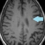
Axial T2
On a T2 weighted image, you can see areas of infarct as bright.
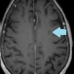
Diffusion
Diffusion weighted imaging is great at detecting areas of infarct.
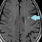
Axial T2*
A T2* is great at identifying is an infarct is hemorrhagic or not.
Acute Infarct
Diffusion Tracer
If an area of infarct is bright on a diffusion weighted image, it can be an acute stroke or potentially T2 shine through artifact.
ADC Map
If the area seen on the diffusion weighted image is dark, it can represent an acute infarct. If it is bright, it is T2 shine through artifact.
Chronic Infarct
Diffusion Tracer
If the area of infarct is dark on the diffusion weighted image, it can be a chronic infarct.
ADC Map
If the area of infarct is bright on the diffusion weighted image, it can represent a chronic infarct or be T2 shine through artifact.
Symptoms
Headache, numbness, weakness in face or arms or legs (usually on one side), confusion, trouble speaking, difficulty with vision, and dizzy.
Treatment
A CT is typically performed first to identify if the stroke is caused from a ruptured vessel. If it is not, tPA can be given to the patient.